
Finding cancer early, when it’s small and before it has spread, often allows for more treatment options. Some early cancers may have signs and symptoms that can be noticed, but that’s not always the case.
CAN GALLBLADDER CANCER BE FOUND EARLY?
Gallbladder cancer is hard to find early (when it’s small and only in the gallbladder). The gallbladder is deep inside the body, so early tumors can’t be seen or felt during routine physical exams. There are no blood tests or other tests that can reliably detect gallbladder cancers early enough to be useful as screening tests. (Screening is testing for cancer in people without any symptoms.) Because of this, most gallbladder cancers are found only after the cancer has grown enough to cause signs or symptoms.
Still, some gallbladder cancers are found before they have spread to other tissues and organs. Many of these early cancers are found unexpectedly when a person’s gallbladder is removed because of gallstones. When the removed gallbladder is looked at in the lab, small cancers or pre-cancers that didn’t cause any symptoms are sometimes found.
SIGNS AND SYMPTOMS OF GALLBLADDER CANCER
Gallbladder cancer doesn’t usually cause signs or symptoms until later in the course of the disease, when the tumor is large and/or has spread. But sometimes symptoms can appear sooner and lead to an early diagnosis. If the cancer is found at an earlier stage, treatment might work better.
Some of the more common symptoms of gallbladder cancer include:
Abdominal (belly) pain
Most people with gallbladder cancer will have belly pain. Most often it’s in the upper right part of the belly.
Nausea and/or vomiting
Some people with gallbladder cancer sometimes have vomiting as a symptom.
Jaundice
If the cancer gets big enough to block the bile ducts, bile from the liver can’t drain into the intestines. This can cause a greenish-yellow chemical (called bilirubin) in the bile to build up in the blood and settle in different parts of the body. The yellow coloring of jaundice can often be seen in the skin and the white part of the eyes.
Lumps in the belly
If the cancer blocks the bile ducts, the gallbladder can swell. Gallbladder cancer can also spread to nearby parts of the liver. These changes can sometimes be felt by the doctor as lumps on the right side of the belly. They can also be seen on imaging tests such as an ultrasound.
Other symptoms
Less common symptoms of gallbladder cancer include:
- Loss of appetite
- Weight loss
- Swelling in the abdomen (belly)
- Fever
- Itchy skin
- Dark urine
- Light-colored or greasy stools
Keep in mind: Gallbladder cancer is rare. These symptoms are more likely to be caused by something other than gallbladder cancer. For example, people with gallstones have many of these same symptoms. And there are many far more common causes of belly pain than gallbladder cancer. Also, hepatitis (liver inflammation caused by a viral infection) is a much more common cause of jaundice.
Still, if you have any of these problems, it’s important to see a doctor right away so the cause can be found and treated, if needed.
TESTS FOR GALLBLADDER CANCER
Some gallbladder cancers are found after the gallbladder has been removed because of gallstones or to treat chronic (long-term) inflammation. Gallbladders removed for those reasons are always looked at under a microscope to see if there’s cancer cells in them.
Most gallbladder cancers, though, aren’t found until a person goes to a doctor because they have symptoms.
Medical history and physical exam
If there’s reason to suspect you might have gallbladder cancer, your doctor will want to take a complete medical history to check for risk factors and to learn more about your symptoms.
Your doctor will examine you to look for signs of gallbladder cancer and other health problems. The exam will focus mostly on the abdomen (belly) to check for any lumps, tenderness, or fluid build-up. The skin and the white part of the eyes will be checked for jaundice (a yellowish color). Sometimes, cancer of the gallbladder spreads to lymph nodes, causing a lump that can be felt beneath the skin. Lymph nodes above the collarbone and in several other locations may be checked.
If symptoms and/or the physical exam suggest you might have gallbladder cancer, tests will be done. These might include lab tests, imaging tests, and other procedures.
Blood tests
Tests of liver and gallbladder function
Lab tests might be done to find out how much bilirubin is in your blood. Bilirubin is the chemical that causes jaundice. Problems in the gallbladder, bile ducts, or liver can raise the blood level of bilirubin.
The doctor may also do tests for albumin, liver enzymes (alkaline phosphatase, AST, ALT, and GGT), and certain other substances in your blood. These may be calledliver function tests. They can help diagnose liver, bile duct, or gallbladder disease.
Tumor markers
Tumor markers are substances made by cancer cells that can sometimes be found in the blood. People with gallbladder cancer may have high blood levels of the markers called CEAand CA 19-9. Usually the blood levels of these markers are high only when the cancer is in an advanced stage . These markers are not specific for gallbladder cancer – that is, other cancers or even some other health conditions also can make them go up.
These tests can sometimes be useful after a person is diagnosed with gallbladder cancer. If the levels of these markers are found to be high, they can be followed over time to help tell how well treatment is working.
Imaging tests
Imaging tests use x-rays, magnetic fields, or sound waves to create pictures of the inside of your body. Imaging tests can be done for a number of reasons, including:
- To look for suspicious areas that might be cancer
- To help a doctor guide a biopsy needle into a suspicious area to take a sample for testing
- To learn how far cancer has spread
- To help make treatment decisions
- To help find out if treatment is working
- To look for signs of the cancer coming back after treatment
People who have (or might have) gallbladder cancer may have one or more of these tests:
Ultrasound
Ultrasound uses sound waves and their echoes to create images of the inside of the body. A small instrument called a transducergives off sound waves and picks up their echoes as they bounce off organs inside the body. The echoes are converted by a computer into an image on a screen.
Abdominal ultrasound: This is often the first imaging test done in people who have symptoms like jaundice or pain in the right upper part of their abdomen (belly). This is an easy test to have and it doesn’t use radiation. You simply lie on a table while a technician moves the transducer on the skin over the right upper abdomen.
This type of ultrasound can also be used to guide a needle into a suspicious area or lymph node so that cells can be removed (biopsied) and looked at under a microscope. This is called an ultrasound-guided needle biopsy.
Endoscopic or laparoscopic ultrasound: In these techniques, the doctor puts the ultrasound transducer inside the body and closer to the gallbladder. This gives more detailed images than a standard ultrasound. The transducer is on the end of a thin, lighted tube that has a camera on it. The tube is either passed through the mouth, down through the stomach, and near the gallbladder (endoscopic ultrasound) or through a small surgical cut on your belly (laparoscopic ultrasound).
If there’s a tumor, ultrasound might help the doctor see if and how far it has spread into the gallbladder wall, which helps in planning for surgery. Ultrasound may be able to show if nearby lymph nodes are enlarged, which can be a sign that cancer has reached them.
Computed tomography (CT) scan
A CT scan uses x-rays to make detailed cross-sectional images of your body. It can be used to
- Help diagnose gallbladder cancer by showing tumors in the area.
- Help stage the cancer (find out how far it has spread). CT scans can show the organs near the gallbladder (especially the liver), as well as lymph nodes and distant organs the cancer might have spread to.
- A type of CT known as CT angiography can be used to look at the blood vessels near the gallbladder. This can help determine if surgery is an option.
- Guide a biopsy needle into a suspected tumor. This is called a CT-guided needle biopsy. To do it, you stay on the CT scanning table while the doctor advances a biopsy needle through your skin and toward the mass. CT scans are repeated until the needle is inside the mass. A small amount of tissue (a sample) is then taken out through the needle.
Magnetic resonance imaging (MRI) scan
Like CT scans, MRI scans show detailed images of soft tissues in the body. But MRI scans use radio waves and strong magnets instead of x-rays. A contrast material called gadolinium may be injected into a vein before the scan to see details better.
MRI scans provide a great deal of detail and can be very helpful in looking at the gallbladder and nearby bile ducts and other organs. Sometimes they can help tell a benign (non-cancer) tumor from one that’s cancer. Special types of MRI scans can also be used in people who may have gallbladder cancer:
- MR cholangiopancreatography (MRCP) can be used to look at the bile ducts and is described below in the section on cholangiography.
- MR angiography (MRA) looks at blood vessels and is also covered in the next section on angiography..
Cholangiography
A cholangiogram is an imaging test that looks at the bile ducts to see if they are blocked, narrowed, or dilated (widened). This can help show if someone might have a tumor that’s blocking a duct. It can also be used to help plan surgery. There are several types of cholangiograms, each of which has different pros and cons.
Magnetic resonance cholangiopancreatography (MRCP): This is a way to get images of the bile ducts using the same type of machine used for standard MRIs. Neither an endoscope or an IV contrast material is used, unlike other types of cholangiograms. Because it’s non-invasive (nothing is put in your body), doctors often use MRCP if they just need images of the bile ducts. This test can’t be used to get biopsy samples of tumors or to place stents (small tubes) in the ducts to keep them open.
Endoscopic retrograde cholangiopancreatography (ERCP):In this procedure, a doctor passes a long, flexible tube (endoscope) down your throat, through your stomach, and into the first part of the small intestine. This is usually done while you are sedated (given medicine to make you sleepy). A small catheter (tube) is passed out of the end of the endoscope and into the common bile duct. A small amount of contrast dye is injected through the catheter. The dye helps outline the bile ducts and pancreatic duct as x-rays are taken. The images can show narrowing or blockage of these ducts. This test is more invasive than MRCP, but it has the advantage of allowing the doctor to take samples of cells or fluid for testing. ERCP can also be used to put a stent (a small tube) into a duct to help keep it open.
Percutaneous transhepatic cholangiography (PTC): To do this procedure, the doctor puts a thin, hollow needle through the skin of your belly and into a bile duct inside the liver. You will get medicine through an IV line to make you sleepy before the test. A local anesthetic is also used to numb the area before putting the needle. A contrast dye is then injected through the needle, and x-rays are taken as it passes through the bile ducts. Like ERCP, this test can also be used to take samples of fluid or tissues or to put a stent (small tube) into a duct to help keep it open. Because it’s more invasive, PTC is not usually used unless ERCP has already been tried or can’t be done for some reason.
Angiography
Angiography or an angiogram is an x-ray test used to look at blood vessels. A thin plastic tube called a catheter is threaded into an artery and a small amount of contrast dye is injected to outline blood vessels. Then x-rays are taken. The images show if blood flow in an area is blocked anywhere or affected by a tumor, as well as any abnormal blood vessels in the area. The test can also show if a gallbladder cancer has grown through the walls of certain blood vessels. This information is mainly used to help surgeons decide whether a cancer can be removed and to help plan the operation.
Angiography can also be done with a CT scan (CT angiography) or an MRI (MR angiography). These tend to be used more often because they give information about the blood vessels without the need for a catheter. You may still need an IV line so that a contrast dye can be injected into the bloodstream during the imaging.
Laparoscopy
Laparoscopy is a type of surgery. The doctor puts a thin tube with a light and a small video camera on the end (a laparoscope) into a small incision (cut) in the front of your abdomen (belly) to look at the gallbladder, liver, and other nearby organs and tissues. (Sometimes more than one cut is made.) This is usually done in the operating room while drugs are used to put you into a deep sleep and not feel pain (general anesthesia) during the surgery.
Laparoscopy can help doctors plan surgery or other treatments, and can help determine the stage (extent) of the cancer. If needed, doctors can also put special instruments in through the incisions to take out biopsy samples for testing.
Laparoscopy is often used to take out your gallbladder. This operation is called alaparoscopic cholecystectomy. If gallbladder cancer is found or suspected during that operation, surgeons usually change to an open cholecystectomy (removal of the gallbladder through a larger cut in the abdomen). The open method lets the surgeon see more and may lower the chance of releasing cancer cells into the abdomen when the gallbladder is removed. The use of the open procedure depends on the size of the cancer and whether surgery can remove it all.
Biopsy
During a biopsy, the doctor removes a tissue sample to be looked at with a microscope to see if cancer (or some other disease) is present. For most types of cancer, a biopsy is needed to make a diagnosis. Biopsies are also used to help find out how far the cancer has spread. This is important when choosing the best treatment plan.
But a biopsy isn’t always done before surgery to remove a gallbladder tumor. Doctors are often concerned that sticking a needle into the tumor or otherwise disturbing it without completely removing it might allow cancer cells to spread.
If imaging tests show a tumor in the gallbladder and there are no clear signs that it has spread, the doctor may decide to proceed directly to surgery and treat the tumor as a gallbladder cancer. (See Surgery for Gallbladder Cancer.) In this case, the gallbladder is checked for cancer after it’s been removed.
In other cases, a doctor may feel that a biopsy of a suspicious area in the gallbladder is the best way to know for sure if it’s cancer. For example, imaging tests may show that a tumor has spread or grown too large to be removed completely by surgery. Many gallbladder cancers are not removable by the time they’re first found.
Types of biopsies
There are many ways to take biopsy samples of the gallbladder.
During cholangiography: If ERCP or PTC is being done, a sample of bile may be collected during the procedure to look for cancer cells in the fluid.
During laparoscopy: As noted earlier, biopsy samples can be taken during laparoscopy. Laparoscopy lets the doctor see the surface of the gallbladder and nearby areas and then take small pieces of tissue from any suspicious areas.
Needle biopsy: If the cancer is too big or has spread to much to be removed with surgery, a needle biopsy may be done to confirm the diagnosis and help guide treatment. For this test, a thin, hollow needle is put in through the skin and into the tumor without making a cut in the skin. (The skin is numbed first with a local anesthetic.) The needle is usually guided into place using ultrasound or CT scans. When the images show that the needle is in the tumor, cells and/or fluid are drawn into the needle and sent to the lab to be tested.
In most cases, this is done as a fine needle aspiration (FNA) biopsy, which uses a very thin needle attached to a syringe to suck out (aspirate) a sample of cells.Sometimes, the FNA doesn’t get enough cells for a definite diagnosis, so a core needle biopsy, which uses a slightly larger needle to get a bigger sample, may be done.
GALLBLADDER CANCER STAGES
After a cancer diagnosis, staging provides important information about the extent of cancer in the body and the likely response to treatment.
Stages
After a person is diagnosed with gallbladder cancer, doctors will try to figure out if it has spread, and if so, how far. This process is called staging. The stage of a cancer describes how much cancer is in the body. It helps determine how serious the cancer is and how best to treat it. Doctors also use a cancer’s stage when talking about survival statistics.
The earliest stage gallbladder cancers (called carcinoma in situ) are stage 0. Stages then range from stages I (1) through IV (4). As a rule, the lower the number, the less the cancer has spread. A higher number, such as stage IV, means cancer has spread more. And within a stage, an earlier letter means a lower stage.
Although each person’s cancer experience is unique, cancers with similar stages tend to have a similar outlook and are often treated in much the same way.
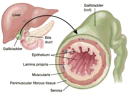
Nearly all gallbladder cancers start in the epithelium (the inside wall of the gallbladder). Over time they grow through the various layers toward the outside of the gallbladder. They may also grow to fill up some or all the space inside the gallbladder at the same time.
How is the stage determined?
The staging system most often used for gallbladder cancer is the American Joint Committee on Cancer (AJCC) TNM system, which is based on 3 key pieces of information:
- The extent (size) of the tumor (T): How far has the cancer grown into the wall of the gallbladder? Has the cancer grown through the gallbladder wall into nearby organs such as the liver?
The gallbladder wall has several layers. From the inside out, these are:- The epithelium, a thin sheet of cells that line the inside wall of the gallbladder
- The lamina propria, a thin layer of loose connective tissue (the epithelium plus the lamina propria form the mucosa)
- The muscularis, a layer of muscular tissue that helps the gallbladder contract, squirting its bile into the bile duct
- The perimuscular (around the muscle) fibrous tissue, another layer of connective tissue
- The serosa, the outer covering of the gallbladder that comes from the peritoneum, which is the lining of the abdominal cavity
The depth that a tumor grows from the inside (epithelium layer) through the other outer layers (all the way through the serosa) is a key part of staging.
- The spread to nearby lymph nodes (N): Has the cancer spread to nearby lymph nodes and if so, how many?
- The spread (metastasis) to distant sites (M): Has the cancer spread to distant organs such as the liver, the peritoneum (the lining of the abdominal cavity), or the lungs?
The system described below is the most recent AJCC system, effective January 2018. This system is used to stage cancers of the gallbladder as well as cancers that start in the cystic duct (the tube that carries bile away from the gallbladder).
The gallbladder staging system uses the pathologic stage(also called thesurgical stage) which is determined by examining the tissue removed during an operation. Sometimes, if surgery can’t be done right away or at all, the cancer will be given a clinical stageinstead. This is based on the results of a physical exam, biopsy, and imaging tests. The clinical stage will be used to help plan treatment. Sometimes, though, the cancer has spread further than the clinical stage estimates, and may not predict the patient’s outlook as accurately as a pathologic stage.
Numbers or letters after T, N, and M provide more details about each of these factors. Higher numbers mean the cancer is more advanced.
Once a person’s T, N, and M categories have been determined, this information is combined in a process called stage grouping to assign an overall stage. For more on this see Cancer Staging.
Cancer staging can be complex, so ask your doctor to explain it to you in a way you understand.
OTHER PROGNOSTIC FACTORS
Besides your stage, there are other factors that can affect your prognosis (outlook).
Grade
The grade describes how closely the cancer cells look like normal gallbladder cells when seen with a microscope.
The scale used for grading gallbladder cancer is from 1 to 3.
- Grade 1 (G1) means the cancer cells look a lot like normal gallbladder cells.
- Grade 3 (G3) means the cancer cells looks very abnormal.
- Grade 2 (G2) falls somewhere in between.
Low-grade cancers (G1) tend to grow and spread more slowly than high-grade (G3) cancers. Most of the time, the outlook is better for Grade 1 and Grade 2 cancers than it is for Grade 3 cancers of the same stage for gallbladder cancer.
Subtype
The specific type of gallbladder cancer you have can influence your outlook. Rare cancer types such as squamous and adenosquamous carcinomas of the gallbladder tend to have a worse prognosis (outlook) than adenocarcinomas (the most common type) and papillary carcinomas.
Lymphovascular Invasion
If cancer cells are seen in small blood vessels (vascular) or lymph vessels (lymphatics) under the microscope, it’s called lymphovascular invasion. When cancer is growing in these vessels, there’s a greater chance that it has spread outside the gallbladder. Gallbladder cancers with lymphovascular invasion tend to have a poor prognosis.
Extent of Resection
If the entire gallbladder tumor can be removed with surgery, it can impact the overall outlook. Cancers that can be removed completely by surgery tend to have a better outlook than those that cannot.
- Resectable cancers are those that doctors believe can be removed completely by surgery.
- Unresectable cancers have spread too far or are in too difficult a place to be removed entirely by surgery.
Only a small percentage of gallbladder cancers are resectable when they’re first found.
QUESTIONS TO ASK ABOUT GALLBLADDER CANCER
It’s important to have honest, open discussions with your cancer care team. They want to answer all of your questions, no matter how minor they might seem. Don’t be afraid to ask them. Here are some questions to get you started:
- Has my cancer spread beyond the gallbladder?
- What’s the stage of my cancer, and what does that mean in my case?
- Do I need other tests before we consider treatment options?
- Do I need to see any other kinds of doctors?
- How much experience do you have treating this type of cancer?
- Should I get a second opinion?
- What are my treatment options?
- Can my cancer be removed with surgery?
- What do you recommend and why?
- What is the goal of treatment?
- What risks or side effects are there to the treatments you suggest? How long are they likely to last?
- How quickly do we need to decide on treatment?
- What should I do to be ready for treatment?
- How long will treatment last? What will it be like? Where will it be done?
- How will treatment affect my daily activities?
- What are the chances my cancer can be cured with these treatment plans?
- What would my options be if the treatment doesn’t work or if the cancer comes back?
- What type of follow-up might I need after treatment?
- Where can I get more information and support?
Along with these, be sure to write down some questions of your own. For instance, you might want more information about recovery times so you can plan your work or activity schedule. Or you might want to ask about qualifying for clinical trials.
Keep in mind that doctors are not the only ones who can provide you with information. Other health care professionals, such as nurses and social workers, may have the answers to some of your questions. You can find out more about speaking with your health care team in The Doctor-Patient Relationship.
Source: American Cancer Society, Gallbladder Cancer, https://www.cancer.org/cancer/gallbladder-cancer.html, July 12, 2018